
The Analytical Chemistry Gap in Drug Quality Control
Gold nanoparticles as colorimetric chemosensors are transforming pharmaceutical quality control — delivering trace-level analytical sensitivity without laboratory infrastructure.The integrity of a pharmaceutical product rests on a single, non-negotiable requirement: that every tablet, capsule, or injectable preparation contains precisely the active pharmaceutical ingredient (API) it claims to contain — at exactly the stated concentration, free from degradation products and contaminants. Verifying this requirement is the domain of pharmaceutical quality control (QC) analysis, and it sits at the heart of patient safety worldwide.
Conventional analytical methods for QC analysis — High-Performance Liquid Chromatography (HPLC), Liquid Chromatography-Mass Spectrometry (LC-MS/MS), Gas Chromatography-Mass Spectrometry (GC-MS), and UV-spectrophotometry — are powerful, sensitive, and rigorously validated. They are also expensive, slow, laboratory-bound, and entirely inaccessible to the majority of pharmaceutical manufacturers and regulatory bodies operating in low- and middle-income countries.
This analytical access gap has real consequences. Substandard and falsified medicines — drugs containing incorrect API concentrations — cause an estimated 169,000 child deaths annually from pneumonia alone, according to the World Health Organization. Rapid, low-cost, field-deployable analytical tools are not a scientific luxury. They are a global health imperative.
Polymer-functionalized metal nanoprobes operating as colorimetric chemosensors represent one of the most promising answers to this challenge — delivering trace-level analytical sensitivity in a format that requires no sophisticated instrumentation, no trained laboratory operator, and no complex sample preparation workflow.
This article provides a comprehensive examination of the science, methodology, performance characteristics, and real-world applications of these nanosensor platforms, with particular focus on their application to antibiotic detection — specifically cephalosporin-class antibiotics — in pharmaceutical formulations and environmental water samples.
1. Foundations — Gold Nanoparticles as Analytical Nanoprobes
1.1 Why Gold?
Among all metallic nanomaterials available to the analytical chemist, gold nanoparticles (AuNPs) occupy a uniquely privileged position. Their analytical utility derives from a quantum optical phenomenon called Localized Surface Plasmon Resonance (LSPR) — the collective oscillation of conduction-band electrons at the nanoparticle surface in response to incident electromagnetic radiation.
For spherical AuNPs in the 10–50 nm diameter range, this resonance condition is met at approximately 520 nm in the visible spectrum — producing the characteristic wine-red coloration of colloidal gold dispersions that has been recognized since Michael Faraday’s pioneering work in 1857. This LSPR absorption peak is extraordinarily sensitive to the nanoparticle’s local environment: changes in particle size, shape, surface chemistry, inter-particle distance, and dielectric properties of the surrounding medium all produce measurable, often visible shifts in the SPR band position and intensity.
This sensitivity is the analytical foundation of colorimetric chemosensing. When a target analyte interacts with the AuNP surface — through direct binding, displacement of stabilizing surface ligands, or charge-mediated aggregation — the resulting change in inter-particle distance shifts the SPR band from ~520 nm toward longer wavelengths (600–700+ nm), producing a color transition from wine-red → purple → blue-grey that is detectable by the naked eye and precisely quantifiable by UV-Vis spectrophotometry.
1.2 Analytical Advantages of AuNP-Based Chemosensors
The analytical profile of AuNP-based colorimetric chemosensors offers several decisive advantages over conventional QC methods:
- Sensitivity: Detection limits routinely reach the micromolar (μM) to nanomolar (nM) range — competitive with HPLC for many pharmaceutical applications
- Speed: Most colorimetric assays yield results within 2–15 minutes of analyte addition, compared to 30–90 minutes for HPLC analysis
- Cost: Reagent costs per analysis are typically 10–50× lower than chromatographic methods
- Simplicity: No mobile phase preparation, no column equilibration, no gradient programming — just mix, observe, measure
- Portability: UV-Vis spectrophotometers are compact and affordable; smartphone-based colorimetric readers eliminate even this requirement
- Naked-eye detection: Qualitative detection requires no instrumentation whatsoever — the color change is directly observable
2. Polymer-Stabilized Gold Nanoparticles as Colorimetric Chemosensors — Engineering Selectivity and Stability
2.1 The Stabilization Challenge
Bare, unfunctionalized gold nanoparticles are thermodynamically unstable in aqueous solution. Without surface stabilization, inter-particle van der Waals attractive forces drive spontaneous irreversible aggregation — collapsing the colloidal dispersion and destroying analytical utility. Effective colorimetric chemosensing requires nanoparticles that are:
- Stable in the absence of the target analyte — maintaining the wine-red baseline color
- Specifically responsive to the target analyte — aggregating selectively upon analyte interaction
- Resistant to non-specific aggregation from interfering matrix components — salts, proteins, competing drug molecules
Polymer surface functionalization addresses all three requirements simultaneously.
2.2 Polyethylene Glycol (PEG) as a Stabilizing and Sensing Agent
Polyethylene glycol (PEG) — a water-soluble, biocompatible, non-toxic polymer of repeating ethylene oxide units [−OCH₂CH₂−]ₙ — is among the most widely employed stabilizing agents for pharmaceutical-grade nanoparticle systems. Its analytical utility as a chemosensor surface coating derives from several complementary properties:
Steric stabilization: PEG chains at the nanoparticle surface create a hydrophilic steric barrier that repels non-specific aggregation from ionic species and biological matrix components. This ensures the nanoparticle dispersion remains stable in complex pharmaceutical and environmental matrices.
Selective molecular interactions: The ether oxygen atoms along the PEG backbone participate in non-covalent interactions — hydrogen bonding, dipole-dipole interactions, and van der Waals forces — with target drug molecules bearing complementary functional groups. These interactions perturb the PEG corona architecture, altering inter-particle distances and triggering the SPR band shift.
Biocompatibility: PEG-coated nanoparticles exhibit minimal cytotoxicity and reduced immunogenicity — critical considerations for any analytical platform intended for pharmaceutical applications.
One-step formulations of PEGylated gold nanoparticles (PEG-AuNPs) have been reported using PEG with different molecular weights as both the reducing and stabilizing agents — simplifying synthesis considerably and eliminating the need for separate reduction and functionalization steps.
2.3 Other Polymer Stabilizers — A Comparative Overview
The polymer-functionalized AuNP chemosensor literature encompasses a diverse range of polymer stabilizing agents, each conferring distinct selectivity profiles:
Polypropylene glycol (PPG): PPG-stabilized AuNPs have been employed as colorimetric probes for third-generation cephalosporin antibiotics, including ceftriaxone, permitting efficient, selective, quantitative, and rapid recognition in a concentration range of 0.1–100 μM in the presence of numerous other drugs and salts.
In a related study, polypropylene glycol was used for stabilization of gold nanoparticles in a simple one-pot two-phase process and subsequently employed for the specific detection of cephradine (CPH) — a first-generation cephalosporin antibiotic. The SPR band was quenched by addition of CPH in the presence of several interferents, and the developed sensor demonstrated potential for fast scanning of pharmaceutical formulations for quantification at production facilities.
Poly(lactic acid) (PLA): PLA-stabilized AuNPs have been developed as a chemosensor for methamidophos — an insecticide — achieving a detection limit of 0.0027 μM, a quantification limit of 0.005 μM, and a linear dynamic range of 0.005–1000 μM, with the presence of interfering insecticides, metal salts, and drugs producing no pronounced effect on quantitative detection.
Polyethylene glycol (PEG) with molecular weight variants: PEG-AuNPs have demonstrated colorimetric sensing capability for biogenic amines, with red-shifting and decrease in SPR peak absorbance along with appearance of an additional peak at ~690 nm observed upon analyte addition — a spectroscopic signature characteristic of AuNP aggregation.
3. The Colorimetric Chemosensor Mechanism — Step by Step
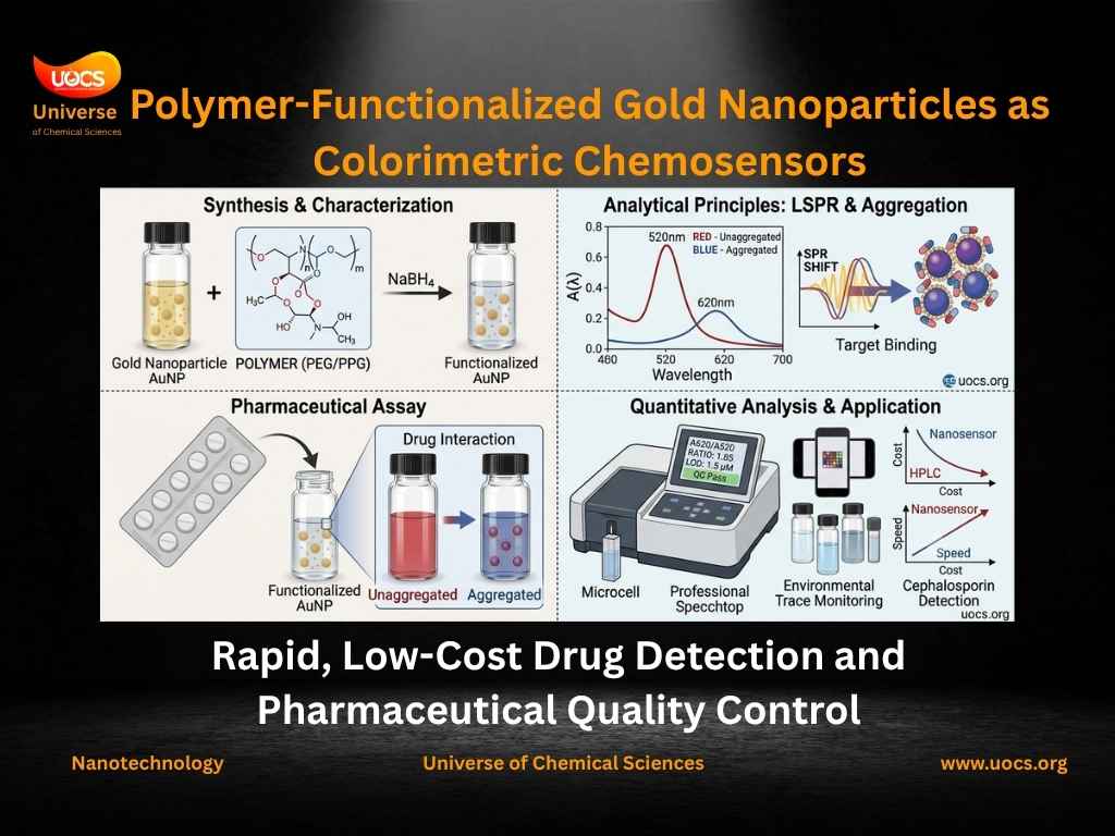
3.1 Synthesis of Polymer-Functionalized AuNPs
The one-pot two-phase chemical reduction method is the most widely employed synthesis approach for polymer-stabilized AuNP chemosensors. The general procedure involves:
Step 1 — Preparation of gold precursor solution: Chloroauric acid (HAuCl₄) is dissolved in deionized water at a defined concentration, typically 1–5 mM. HAuCl₄ serves as the Au³⁺ ion source.
Step 2 — Addition of reducing agent: A chemical reducing agent — most commonly sodium borohydride (NaBH₄) or trisodium citrate — is added to reduce Au³⁺ → Au⁰. The reduction is rapid and exothermic, with immediate color development signaling nanoparticle nucleation.
Step 3 — Polymer stabilization: The polymer stabilizing agent (PEG, PPG, PLA, etc.) is introduced simultaneously with or immediately following reduction. Polymer chains adsorb to the nascent AuNP surface through non-covalent interactions, terminating particle growth and establishing the steric stabilization corona.
Step 4 — Purification and characterization: The resulting polymer-AuNP colloid is characterized by:
- UV-Vis spectrophotometry — confirms SPR band position (~520 nm) and intensity
- Atomic Force Microscopy (AFM) — determines particle size, size distribution, and morphology
- Fourier Transform Infrared Spectroscopy (FTIR) — confirms polymer surface functionalization through characteristic vibrational signatures
- Dynamic Light Scattering (DLS) / Zetasizer — measures hydrodynamic diameter and zeta potential (colloidal stability indicator)
3.2 The Sensing Event — Molecular Mechanism
The colorimetric sensing event proceeds through the following molecular sequence:
Phase 1 — Baseline establishment: Polymer-AuNPs dispersed in aqueous solution display a sharp, well-defined SPR absorption band at ~520 nm. The colloid appears wine-red. This is the analytical baseline.
Phase 2 — Analyte introduction: The target drug molecule (e.g., cephalexin, ceftriaxone, cephradine) is introduced to the AuNP dispersion. The drug molecule interacts with the polymer corona through:
- Hydrogen bonding between drug functional groups (−NH₂, −COOH, −OH) and polymer ether oxygens
- Electrostatic interactions between charged drug moieties and the nanoparticle surface
- Competitive displacement of polymer chains from nanoparticle surface binding sites
Phase 3 — Aggregation cascade: Analyte-mediated perturbation of the protective polymer corona reduces inter-particle electrostatic repulsion, allowing van der Waals attractive forces to drive controlled aggregation. AuNPs cluster into larger assemblies.
Phase 4 — SPR band shift: As inter-particle distances decrease in aggregated clusters, electromagnetic coupling between adjacent nanoparticles red-shifts the collective SPR band from ~520 nm toward 600–700+ nm. This shift is:
- Bathochromic (toward longer wavelengths)
- Hyperchromic at the new peak wavelength
- Hypochromic at the original 520 nm peak
Phase 5 — Color transition: The spectroscopic changes manifest as a visible color transition: wine-red → purple → blue-grey — directly observable to the naked eye and precisely quantifiable by measuring the absorbance ratio A₆₂₀/A₅₂₀ or the shift in λmax.
4. Analytical Performance — Qualitative and Quantitative Analysis
4.1 Qualitative Detection — Naked-Eye Screening
The most immediate application of polymer-AuNP colorimetric chemosensors is rapid qualitative screening — determining the presence or absence of a target drug above a defined threshold concentration without any instrumentation.
This capability is directly actionable for:
- Counterfeit drug detection at point-of-entry customs inspections
- Field-based environmental screening of water sources near pharmaceutical manufacturing facilities
- Rapid lot acceptance/rejection decisions at pharmaceutical receiving docks in resource-limited settings
The binary color change — red (drug absent) versus blue/purple (drug present) — requires no training, no equipment, and yields results in under 5 minutes.
4.2 Quantitative Analysis — Calibration and Linear Dynamic Range
For precise API concentration determination, polymer-AuNP chemosensors are used in quantitative colorimetric mode, with UV-Vis spectrophotometric measurement of the SPR band shift across a calibrated concentration range.
Calibration procedure:
- Prepare a series of drug standard solutions across the expected concentration range
- Add each standard to the polymer-AuNP dispersion under standardized conditions (fixed volume, pH, temperature, reaction time)
- Record UV-Vis absorbance spectra for each concentration
- Plot absorbance ratio (A₆₂₀/A₅₂₀) or Δλmax versus drug concentration
- Fit the linear portion of the response curve to generate the calibration equation
Typical analytical performance parameters for polymer-AuNP colorimetric chemosensors:
| Parameter | Typical Range |
|---|---|
| Linear detection range | 0.01–120 mM (varies by system) |
| Limit of Detection (LOD) | 0.003–11 μM |
| Limit of Quantification (LOQ) | 0.005–33 μM |
| Correlation coefficient (R²) | >0.99 |
| Analysis time | 2–15 minutes |
| Sample volume | 0.1–3 mL |
4.3 Selectivity — The Critical Analytical Challenge
Pharmaceutical and environmental samples are chemically complex matrices containing hundreds of co-existing species — excipients, degradation products, other drugs, metal ions, biological macromolecules. A colorimetric chemosensor with poor selectivity produces false positives that render it analytically useless.
Selectivity evaluation involves systematic investigation of the sensor response to a panel of potential interferents — typically including:
- Other antibiotics of the same and different classes
- Common pharmaceutical excipients (starch, lactose, magnesium stearate)
- Metal ions (Na⁺, K⁺, Ca²⁺, Mg²⁺, Fe³⁺, Cu²⁺)
- Amino acids and proteins
- Environmental matrix components (humic acids, dissolved organic carbon)
Well-designed polymer-AuNP systems demonstrate that the presence of other interfering insecticides, metal salts, and drugs produces no pronounced effect on quantitative target detection — a critical selectivity benchmark for real sample analysis.
The molecular basis of selectivity lies in the complementary functional group geometry between the target drug and the polymer surface. Cephalosporin antibiotics, for example, possess a characteristic β-lactam ring with flanking carboxyl and amino groups that interact specifically with ether-rich polymer coronas in ways that structurally dissimilar molecules cannot replicate.
5. Application to Cephalosporin Antibiotic Detection
5.1 The Cephalosporin Class — Structure and Analytical Relevance
Cephalosporins are a major class of β-lactam antibiotics characterized by a dihydrothiazine ring fused to a β-lactam ring — the 7-aminocephalosporanic acid (7-ACA) core. They are classified into five generations based on their antimicrobial spectrum:
- First generation: Cephalexin, Cephradine, Cefazolin — narrow spectrum, gram-positive coverage
- Second generation: Cefaclor, Cefuroxime — extended gram-negative coverage
- Third generation: Ceftriaxone, Cefotaxime, Ceftazidime — broad spectrum, including resistant organisms
- Fourth generation: Cefepime — extended spectrum including Pseudomonas
- Fifth generation: Ceftaroline — active against MRSA
β-lactams, including cephalosporins, penicillins, carbapenems, and monobactams, represent effective medicines widely used in the treatment of mastitis, pulmonary, urinary, and septicemia conditions, and are also used in boosting animal growth.
Their widespread clinical and veterinary use makes cephalosporins among the most environmentally prevalent pharmaceutical contaminants — detected in hospital effluents, municipal wastewater, river systems, and groundwater at concentrations ranging from nanograms to micrograms per liter.
5.2 Cephalexin — A Primary Target for Colorimetric Detection
Cephalexin (CFX) — a first-generation cephalosporin antibiotic — is one of the most widely prescribed oral antibiotics globally, used for the treatment of respiratory tract infections, urinary tract infections, skin and soft tissue infections, and otitis media. Its chemical structure features:
- A β-lactam ring — the pharmacophoric core responsible for antibacterial activity
- A dihydrothiazine ring — providing structural rigidity
- A phenylglycine side chain at C-7 — contributing to the D-phenylglycyl moiety
- Carboxyl (−COOH) and amino (−NH₂) functional groups — the primary interaction sites with polymer-AuNP surfaces
These functional groups enable specific non-covalent interactions with polymer-stabilized AuNP surfaces — particularly hydrogen bonding and electrostatic interactions — that drive the selective aggregation response underlying colorimetric detection.
PEG-functionalized AuNPs (PEG-AuNPs) have been specifically investigated as colorimetric nanoprobes for cephalexin detection, demonstrating:
- Linear detection range: 0.1–100 μM
- Limit of detection in the low μM range — sufficient for pharmaceutical QC and environmental monitoring applications
- Selective response in the presence of common pharmaceutical excipients and competing drug molecules
- Applicability to real pharmaceutical formulation samples (cephalexin capsules) with recovery values consistent with declared label claims
5.3 Comparative Performance — Polymer-AuNP Sensors vs. Conventional Methods
| Analytical Method | LOD | Analysis Time | Cost per Analysis | Infrastructure Required |
|---|---|---|---|---|
| HPLC | ~0.01 μM | 30–90 min | High | Centralized laboratory |
| LC-MS/MS | ~0.001 μM | 45–120 min | Very high | Specialized laboratory |
| UV-Spectrophotometry | ~1–5 μM | 10–20 min | Low-medium | Basic laboratory |
| Polymer-AuNP Colorimetric | 0.003–11 μM | 2–15 min | Very low | Minimal / field-deployable |
| Aptamer-AuNP Colorimetric | ~0.1 nM | 15–30 min | Low-medium | Basic laboratory |
6. Environmental Monitoring Applications
6.1 Pharmaceutical Contamination of Aquatic Systems
The environmental fate of pharmaceutical APIs following consumption, excretion, and disposal represents one of the most pressing challenges in environmental analytical chemistry. Cephalosporin antibiotics enter aquatic systems through multiple pathways:
- Hospital effluents — the highest-concentration source, with cephalexin detected at 1–100 μg/L in untreated hospital wastewater
- Municipal wastewater treatment plant effluents — conventional biological treatment removes only 20–80% of cephalosporin residues
- Agricultural runoff — veterinary cephalosporin use in livestock contributes to soil and groundwater contamination
- Pharmaceutical manufacturing effluents — process wastewater from API synthesis can contain mg/L concentrations
Water pollution by antibiotics and heavy metals threatens the ecological environment and human health globally, yet there is no rapid method to detect multiple antibiotics and metal ions simultaneously — a gap that polymer-AuNP colorimetric chemosensors are uniquely positioned to address.
6.2 Environmental Detection Performance
For environmental monitoring applications, polymer-AuNP chemosensors must demonstrate reliable performance in complex natural water matrices — river water, groundwater, treated wastewater — containing organic matter, dissolved salts, suspended particles, and competing analytes.
ATG-functionalized AuNPs have been demonstrated as colorimetric chemosensors for the simultaneous detection of fluoroquinolone antibiotics and heavy metal ions in lake and tap water samples, with detection limits of 1.57 μM and 1.30 μM for moxifloxacin and ciprofloxacin respectively, and 57.1 nM for Cr(III).
The spike-and-recovery methodology is standard for validating environmental applicability: known concentrations of target analyte are added to real environmental water samples, and the sensor’s ability to accurately quantify the added concentration — expressed as percentage recovery — is determined. Recovery values of 95–105% are generally considered acceptable for environmental analytical methods.
7. Nanosensor Classification — Understanding the Terminology
The colorimetric chemosensor literature employs several overlapping terms that warrant precise definition:
Nanosensor: The broadest term — any sensing device that incorporates nanoscale materials (1–100 nm) as the active sensing element. All polymer-AuNP colorimetric platforms are nanosensors.
Nanobiosensor: A nanosensor that incorporates a biological recognition element — aptamer, antibody, enzyme, DNA — as the selectivity-conferring component. Aptamer-functionalized AuNP sensors for antibiotic detection are nanobiosensors.
Nanoprobe: Emphasizes the investigative function of the nanoparticle — its role as a molecular-scale probe that reports on its chemical environment. The term is particularly appropriate for AuNP systems used in investigation of complex pharmaceutical and environmental matrices.
Colorimetric chemosensor: Specifies both the transduction mechanism (colorimetry — optical color change) and the recognition basis (chemical interactions — non-biological molecular recognition). Polymer-AuNP platforms are typically classified as colorimetric chemosensors when the recognition element is a synthetic polymer rather than a biological molecule.
Assay: The complete analytical procedure — from sample preparation through measurement and data interpretation — for determining the presence or concentration of a target analyte. A colorimetric assay using polymer-AuNP nanoprobes encompasses the full analytical workflow.
8. Validation Parameters for Pharmaceutical QC Applications
For polymer-AuNP colorimetric methods to be accepted as legitimate pharmaceutical QC tools — either as primary analytical methods or as rapid screening complements to HPLC — they must be validated according to internationally recognized guidelines, including ICH Q2(R2) and USP <1225>.
Key validation parameters include:
Linearity and range: Demonstration that the analytical response (A₆₂₀/A₅₂₀ or Δλmax) is proportional to analyte concentration across the defined linear dynamic range, with R² ≥ 0.999.
Limit of Detection (LOD): The lowest analyte concentration producing a signal distinguishable from baseline noise at defined statistical confidence — calculated as LOD = 3σ/S, where σ is the standard deviation of the blank response and S is the calibration slope.
Limit of Quantification (LOQ): The lowest concentration permitting quantitative determination with acceptable precision and accuracy — calculated as LOQ = 10σ/S.
Precision: Repeatability (intra-day) and intermediate precision (inter-day) expressed as relative standard deviation (RSD%), typically required to be <2% for pharmaceutical applications.
Accuracy: Percentage recovery of known analyte concentrations added to pharmaceutical formulation matrices — acceptable range 98–102% for API determination.
Selectivity: Demonstrated absence of analytical interference from common excipients, degradation products, and co-administered drugs.
Robustness: Stability of analytical results under deliberate small variations in method parameters — pH, temperature, reagent concentration, reaction time.
9. Current Limitations and Future Directions
9.1 Present Limitations
Despite their considerable promise, polymer-AuNP colorimetric chemosensors face several challenges before achieving widespread pharmaceutical and regulatory acceptance:
Matrix complexity: Real pharmaceutical and environmental samples present analytical challenges — protein binding, pH variation, ionic strength effects — that can perturb colorimetric responses and require method-specific optimization.
Multiplexed detection: Multiplex colorimetric aptasensors capable of detecting more than one antibiotic simultaneously have been demonstrated but remain technically complex — most deployed colorimetric platforms target single analytes.
Quantitative precision: The inherent variability of color-based measurements — affected by lighting conditions, observer perception, and smartphone camera calibration — limits quantitative precision compared to HPLC. Standardized colorimetric readers and dedicated smartphone applications are actively being developed to address this.
Regulatory acceptance: No polymer-AuNP colorimetric method has yet achieved formal inclusion in major pharmacopoeias (USP, BP, EP) as an official analytical method — limiting their use to research and rapid screening contexts in regulated pharmaceutical environments.
9.2 Emerging Directions
Smartphone-integrated colorimetric platforms: Coupling polymer-AuNP sensors with smartphone camera-based colorimetric readers enables quantitative analysis without dedicated spectrophotometers — creating truly portable, field-deployable QC tools deployable at the point of need.
Paper-based microfluidic colorimetric assays: Integrating polymer-AuNP sensing chemistry into paper microfluidic substrates (μPADs) creates single-use, disposable test strips analogous to lateral flow immunoassays — with potential for high-throughput pharmaceutical screening at minimal cost.
Machine learning-assisted color analysis: Training convolutional neural networks to quantify colorimetric responses from smartphone images is emerging as a powerful approach to improving quantitative precision and eliminating observer-dependent variability.
Dual-mode sensors: Combining colorimetric with fluorometric or electrochemical transduction in a single nanoparticle platform provides orthogonal analytical confirmation — enhancing reliability in complex matrix applications.
Conclusion: Democratizing Pharmaceutical Quality Control
The polymer-functionalized metal nanoprobe colorimetric chemosensor represents a transformative analytical platform — one that democratizes the sensitivity of laboratory-grade pharmaceutical analysis and places it in a format accessible to quality control professionals, environmental monitors, and regulatory inspectors operating anywhere in the world, regardless of laboratory infrastructure.
From the SPR band shift of aggregating PEG-AuNPs in the presence of cephalexin, to the trace-level limit of detection achieved by optimized polymer-stabilized nanosensor systems, to the selective, quantitative determination of APIs in complex pharmaceutical formulations and contaminated environmental water samples — these platforms embody chemistry’s most powerful promise: making the invisible analytically visible.
As validation frameworks mature, smartphone integration advances, and regulatory acceptance expands, polymer-AuNP colorimetric chemosensors are positioned to become a standard tool in the global pharmaceutical QC toolkit — closing the analytical access gap that currently leaves billions of patients in the developing world exposed to unverified, potentially substandard medicines.
Key Takeaways
- Gold nanoparticles are ideal colorimetric nanoprobes due to their tunable SPR band (~520 nm) and sensitivity to inter-particle distance changes
- Polymer functionalization (PEG, PPG, PLA) confers colloidal stability and analyte-selective recognition through non-covalent surface interactions
- Cephalosporin antibiotics — including cephalexin, cephradine, and ceftriaxone — are primary targets with detection ranges of 0.1–100 μM
- Qualitative detection (naked-eye color change) enables rapid screening; quantitative analysis via UV-Vis spectrophotometry enables precise API determination
- Validation parameters (LOD, LOQ, linearity, selectivity, accuracy, precision) must meet ICH Q2(R2) standards for pharmaceutical QC acceptance
- Environmental applications include detection of antibiotic residues in hospital effluents, river water, and treated wastewater
- Future directions include smartphone integration, paper microfluidics, and machine learning-assisted colorimetric quantification
References:
[1] Raja, D. A.; Munir, F.; Shah, M. R.; Bhanger, M. I.; Malik, M. I. Colorimetric Sensing of Cephradine through Polypropylene Glycol Functionalized Gold Nanoparticles. Royal Society Open Science 2021, 8, 210185.
🔗 https://doi.org/10.1098/rsos.210185
[2] Kusuma, S. A. F.; Harmonis, J. A.; Pratiwi, R.; Hasanah, A. N. Gold Nanoparticle-Based Colorimetric Sensors: Properties and Application in Detection of Heavy Metals and Biological Molecules. Sensors 2023, 23 (19), 8172.
🔗 https://doi.org/10.3390/s23198172
[3] Islam, I. U.; Qurashi, A. N.; Adnan, A. et al. Bioremediation and Adsorption: Strategies for Managing Pharmaceutical Pollution in Aquatic Environment. Water Air and Soil Pollution 2025, 236, 579.
🔗 https://doi.org/10.1007/s11270-025-08193-7
This article is the comprehensive technical companion to the InfoChemist.com explainer: What Is a Colorimetric Sensor? Nanoparticles Detecting Drugs in Water. Read the original news brief at infochemist.com.
Continue your learning journey and Explore Chemistry with interactive topics, guides, and resources across all branches of chemistry.
Discover how innovation is transforming chemistry — explore our detailed guide on Future Technologies in Chemical Sciences to see what’s next in research and industry.
— UOCS Editorial Team | uocs.org — Accelerating Scientific Discovery

